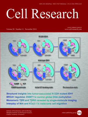
Volume 20, No 11, Nov 2010
ISSN: 1001-0602
EISSN: 1748-7838 2018
impact factor 17.848*
(Clarivate Analytics, 2019)
Volume 20 Issue 11, November 2010: 1216-1223
ORIGINAL ARTICLES
Monomeric type I and type III transforming growth factor-β receptors and their dimerization revealed by single-molecule imaging
Wei Zhang1, Jinghe Yuan1, Yong Yang1, Li Xu1, Qiang Wang2,3, Wei Zuo2, Xiaohong Fang1 and Ye-Guang Chen2
1Beijing National Laboratory for Molecular Sciences, Institute of Chemistry, Key Laboratory of Molecular Nanostructures and Nanotechnology, Chinese Academy of Sciences, Beijing 100190, China
2State Key Laboratory of Biomembrane and Membrane Biotechnology, School of Life Sciences, Tsinghua University, Beijing 100084, China
3Current address: Institute of Zoology, Chinese Academy of Sciences, Beijing 100101, China
Correspondence: Xiaohong Fang, Ye-Guang Chen,(xfang@iccas.ac.cn; ygchen@mail.tsinghua.edu.cn)
Transforming growth factor-β (TGF-β) binds with two transmembrane serine/threonine kinase receptors, type II (TβRII) and type I receptors (TβRI), and one accessory receptor, type III receptor (TβRIII), to transduce signals across cell membranes. Previous biochemical studies suggested that TβRI and TβRIII are preexisted homo-dimers. Using single-molecule microscopy to image green fluorescent protein-labeled membrane proteins, for the first time we have demonstrated that TβRI and TβRIII could exist as monomers at a low expression level. Upon TGF-β1 stimulation, TβRI follows the general ligand-induced receptor dimerization model for activation, but this process is TβRII-dependent. The monomeric status of the non-kinase receptor TβRIII is unchanged in the presence of TGF-β1. With the increase of receptor expression, both TβRI and TβRIII can be assembled into dimers on cell surfaces.
Cell Research (2010) 20:1216-1223. doi:10.1038/cr.2010.105; published online 13 July 2010
FULL TEXT | PDF
Browse 2190


