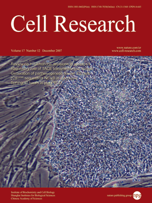
Volume 17, No 12, Dec 2007
ISSN: 1001-0602
EISSN: 1748-7838 2018
impact factor 17.848*
(Clarivate Analytics, 2019)
Volume 17 Issue 12, December 2007: 1008-1019
ORIGINAL ARTICLES
Derivation of human embryonic stem cell lines from parthenogenetic blastocysts
Qingyun Mai1,*, Yang Yu1,2,3,*, Tao Li1, Liu Wang2, Mei-jue Chen4, Shu-zhen Huang4, Canquan Zhou1 and Qi Zhou2
1Reproductive Medical Center, the First Affiliated Hospital of SUMS University, Guangzhou 210029, China
2State Key Laboratory of Reproductive Biology, Institute of Zoology, Chinese Academy of Sciences, Beijing 100101, China
3Graduate University of Chinese Academy of Sciences, Beijing, China
4Ministry of Health Key Lab of Embryo Molecular Biology, Institute of Medical Genetics, Shanghai Jiao Tong University School of Medicine, Shanghai 200040, China
Correspondence: Qi Zhou Canquan Zhou Shu-zhen Huang(qzhou@ioz.ac.cn zhoucanquan@hotmail.com szhuang1@yahoo.com)
Parthenogenesis is one of the main, and most useful, methods to derive embryonic stem cells (ESCs), which may be an important source of histocompatible cells and tissues for cell therapy. Here we describe the derivation and characterization of two ESC lines (hPES-1 and hPES-2) from in vitro developed blastocysts following parthenogenetic activation of human oocytes. Typical ESC morphology was seen, and the expression of ESC markers was as expected for alkaline phosphatase, octamer-binding transcription factor 4, stage-specific embryonic antigen 3, stage-specific embryonic antigen 4, TRA-1-60, and TRA-1-81, and there was absence of expression of negative markers such as stage-specific embryonic antigen 1. Expression of genes specific for different embryonic germ layers was detected from the embryoid bodies (EBs) of both hESC lines, suggesting their differentiation potential in vitro. However, in vivo, only hPES-1 formed teratoma consisting of all three embryonic germ layers (hPES-2 did not). Interestingly, after continuous proliferation for more than 100 passages, hPES-1 cells still maintained a normal 46 XX karyotype; hPES-2 displayed abnormalities such as chromosome translocation after long term passages. Short Tandem Repeat (STR) results demonstrated that the hPES lines were genetic matches with the egg donors, and gene imprinting data confirmed the parthenogenetic origin of these ES cells. Genome-wide SNP analysis showed a pattern typical of parthenogenesis. All of these results demonstrated the feasibility to isolate and establish human parthenogenetic ESC lines, which provides an important tool for studying epigenetic effects in ESCs as well as for future therapeutic interventions in a clinical setting.
Cell Research (2007) 17: 1008-1019. doi: 10.1038/cr.2007.102; published online 11 December 2007
FULL TEXT | PDF
Browse 2076


