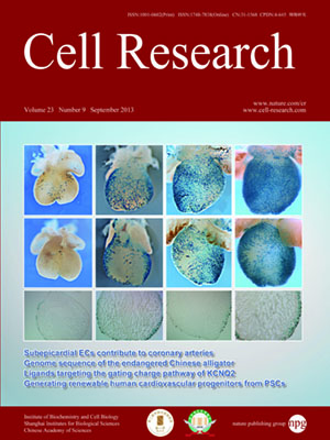
Volume 23, No 9, Sep 2013
ISSN: 1001-0602
EISSN: 1748-7838 2018
impact factor 17.848*
(Clarivate Analytics, 2019)
Volume 23 Issue 9, September 2013: 1075-1090
ORIGINAL ARTICLES
Subepicardial endothelial cells invade the embryonic ventricle wall to form coronary arteries
Xueying Tian1,*, Tianyuan Hu1,*, Hui Zhang1,*, Lingjuan He1, Xiuzhen Huang1, Qiaozhen Liu1, Wei Yu1, Liang He1, Zhongzhou Yang2, Zhen Zhang3, Tao P Zhong4, Xiao Yang5, Zhen Yang6, Yan Yan6, Antonio Baldini7, Yunfu Sun8, Jie Lu9, Robert J Schwartz10, Sylvia M Evans11, Adriana C Gittenberger-de Groot12, Kristy Red-Horse13 and Bin Zhou1
1Key Laboratory of Nutrition and Metabolism, Institute for Nutritional Sciences, Shanghai Institutes for Biological Sciences, Graduate School of the Chinese Academy of Sciences, Chinese Academy of Sciences, Shanghai 200031, China
2Model Animal Research Center of Nanjing University, Nanjing, Jiangsu 210061, China
3Shanghai Children's Medical Center, Shanghai Jiao Tong University School of Medicine, Shanghai 200092, China
4State Key Laboratory of Genetic Engineering, School of Life Sciences, Fudan University, Shanghai 200433, China
5State Key Laboratory of Proteomics, Institute of Biotechnology, Beijing 100071, China
6Zhongshan Hospital, Fudan University, Shanghai 200032, China
7Institute of Genetics and Biophysics CNR, University of Naples Federico II, 80138 Naples, Italy
8School of Medicine, Tongji University, Shanghai 201000, China
9Shanghai 10th People's Hospital, Tongji University, School of Medicine, Shanghai 200072, China
10Department of Biology and Biochemistry, University of Houston, Houston, TX 77204, USA
11The Skaggs School of Pharmacy and Pharmaceutical Science, University of California San Diego, La Jolla, CA 92093, USA
12Department of Cardiology, Leiden University Medical Center, Postal zone S-5-24, PO Box 9600, 2300 RC Leiden, The Netherlands
13Department of Biology, School of Medicine, Stanford University, Stanford, CA 94305, USA
Correspondence: Bin Zhou(zhoubin@sibs.ac.cn)
Coronary arteries bring blood flow to the heart muscle. Understanding the developmental program of the coronary arteries provides insights into the treatment of coronary artery diseases. Multiple sources have been described as contributing to coronary arteries including the proepicardium, sinus venosus (SV), and endocardium. However, the developmental origins of coronary vessels are still under intense study. We have produced a new genetic tool for studying coronary development, an AplnCreER mouse line, which expresses an inducible Cre recombinase specifically in developing coronary vessels. Quantitative analysis of coronary development and timed induction of AplnCreER fate tracing showed that the progenies of subepicardial endothelial cells (ECs) both invade the compact myocardium to form coronary arteries and remain on the surface to produce veins. We found that these subepicardial ECs are the major sources of intramyocardial coronary vessels in the developing heart. In vitro explant assays indicate that the majority of these subepicardial ECs arise from endocardium of the SV and atrium, but not from ventricular endocardium. Clonal analysis of Apln-positive cells indicates that a single subepicardial EC contributes equally to both coronary arteries and veins. Collectively, these data suggested that subepicardial ECs are the major source of intramyocardial coronary arteries in the ventricle wall, and that coronary arteries and veins have a common origin in the developing heart.
10.1038/cr.2013.83
FULL TEXT | PDF
Browse 2931


