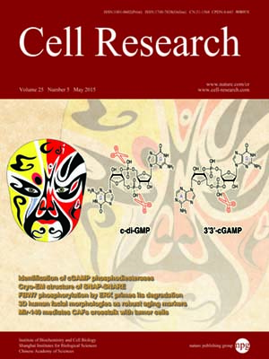
Volume 25, No 5, May 2015
ISSN: 1001-0602
EISSN: 1748-7838 2018
impact factor 17.848*
(Clarivate Analytics, 2019)
Volume 25 Issue 5, May 2015: 551-560
ORIGINAL ARTICLES
Cryo-EM structure of SNAP-SNARE assembly in 20S particle
Qiang Zhou1,*, Xuan Huang1,*, Shan Sun1,*, Xueming Li2, Hong-Wei Wang2 and Sen-Fang Sui1
1State Key Laboratory of Biomembrane and Membrane Biotechnology, Center for Structural Biology, School of Life Sciences, Tsinghua University, Beijing 100084, China
2Ministry of Education Key Laboratory of Protein Science, Center for Structural Biology, Tsinghua-Peking Joint Center for Life Sciences, School of Life Sciences, Tsinghua University, Beijing 100084, China
Correspondence: Sen-Fang Sui; Hong-Wei Wang(suisf@mail.tsinghua.edu.cn; hongweiwang@tsinghua.edu.cn)
N-ethylmaleimide-sensitive factor (NSF) and α soluble NSF attachment proteins (α-SNAPs) work together within a 20S particle to disassemble and recycle the SNAP receptor (SNARE) complex after intracellular membrane fusion. To understand the disassembly mechanism of the SNARE complex by NSF and α-SNAP, we performed single-particle cryo-electron microscopy analysis of 20S particles and determined the structure of the α-SNAP-SNARE assembly portion at a resolution of 7.35 Å. The structure illustrates that four α-SNAPs wrap around the single left-handed SNARE helical bundle as a right-handed cylindrical assembly within a 20S particle. A conserved hydrophobic patch connecting helices 9 and 10 of each α-SNAP forms a chock protruding into the groove of the SNARE four-helix bundle. Biochemical studies proved that this structural element was critical for SNARE complex disassembly. Our study suggests how four α-SNAPs may coordinate with the NSF to tear the SNARE complex into individual proteins.
10.1038/cr.2015.47
FULL TEXT | PDF
Browse 2299


