Advanced Search

Submit Manuscript

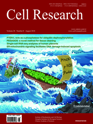
Volume 28, No 8, Aug 2018

ISSN: 1001-0602 
EISSN: 1748-7838 2018 
impact factor 17.848* 
(Clarivate Analytics, 2019)

Volume 28 Issue 8, August 2018: 833-854   |  Open Access

ORIGINAL ARTICLES

DNA damage triggers tubular endoplasmic reticulum extension to promote apoptosis by facilitating ER-mitochondria signaling

Pengli Zheng 1,2, Qingzhou Chen 1, Xiaoyu Tian 1, Nannan Qian 1,3, Peiyuan Chai 1, Bing Liu 1,4, Junjie Hu 5,6, Craig Blackstone 2,Desheng Zhu 7, Junlin Teng 1 and Jianguo Chen 1,4

1 Key Laboratory of Cell Proliferation and Differentiation of the Ministry of Education, State Key Laboratory of Membrane Biology, College of Life Sciences, Peking University, Beijing 100871, China; 2 Cell Biology Section, Neurogenetics Branch, National Institute of Neurological Disorders and Stroke, National Institutes of Health, Bethesda, Maryland 20892, USA; 3 College of Life Sciences, Jiangsu Normal University, Xuzhou 221116, China; 4 Center for Quantitative Biology, Peking University, Beijing 100871, China; 5 National Laboratory of Macromolecules, Institute of Biophysics, Chinese Academy of Science, Beijing 100871, China; 6 Department of Genetics and Cell Biology, College of Life Sciences, Nankai University and Tianjin Key Laboratory of Protein Sciences, Tianjin 300071, China and 7 Laboratory Animal Research Center, Peking University, Beijing 100871, China
Correspondence: Junlin Teng (junlinteng@pku.edu.cn) or Jianguo Chen (chenjg@pku.edu.cn)
Correspondence: Pengli Zheng, Qingzhou Chen, Xiaoyu Tian.

The endoplasmic reticulum (ER) is composed of the nuclear envelope, perinuclear sheets and a peripheral tubular network. The peripheral ER and mitochondria form tight contacts at specific subdomains, which coordinate the functions of the two organelles and are required for multiple cellular processes such as Ca2+ transfer and apoptosis. However, it is largely unknown how ER morphology and ER-mitochondria signaling are dynamically regulated under different physiological or pathological conditions such as DNA damage. Here we show that the peripheral, tubular ER undergoes significant extension in response to DNA damage, and that this process is dependent on p53-mediated transcriptional activation of the ER-shaping proteins REEP1, REEP2 and EI24 (alias PIG8). This promotes the formation of ER-mitochondria contacts through EI24 and the mitochondrial outer membrane protein VDAC2, facilitates Ca2+ transfer from ER to mitochondria and promotes DNA damage-induced apoptosis. Thus, we identify a unique DNA damage response pathway involving alterations in ER morphology, ER-mitochondria signaling, and apoptosis.


https://doi.org/10.1038/s41422-018-0065-z

FULL TEXT | PDF

Browse 1628