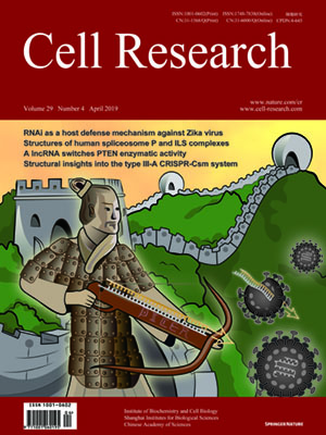
Volume 29, No 4, Apr 2019
ISSN: 1001-0602
EISSN: 1748-7838 2018
impact factor 17.848*
(Clarivate Analytics, 2019)
Volume 29 Issue 4, April 2019: 274-285 | Open Access
ORIGINAL ARTICLES
Structures of the human spliceosomes before and after release of the ligated exon
Xiaofeng Zhang 1, Xiechao Zhan 1, Chuangye Yan 1, Wenyu Zhang 1, Dongliang Liu 1, Jianlin Lei 1,2 and Yigong Shi 1,3
1Beijing Advanced Innovation Center for Structural Biology, Tsinghua-Peking Joint Center for Life Sciences, School of Life Sciences and School of Medicine, Tsinghua University,
Beijing 100084, China; 2Technology Center for Protein Sciences, Ministry of Education Key Laboratory of Protein Sciences, School of Life Sciences, Tsinghua University, Beijing
100084, China and 3School of Life Sciences, Westlake University, 18 Shilongshan Road, Xihu District, Hangzhou, Zhejiang 310024, China
Correspondence: Chuangye Yan (yancy@mails.tsinghua.edu.cn) or Yigong Shi (shi-lab@tsinghua.edu.cn)These authors contributed equally: Xiaofeng Zhang, Xiechao Zhan, Chuangye Yan, Wenyu Zhang
Pre-mRNA splicing is executed by the spliceosome, which has eight major functional states each with distinct composition. Five of these eight human spliceosomal complexes, all preceding exon ligation, have been structurally characterized. In this study, we report the cryo-electron microscopy structures of the human post-catalytic spliceosome (P complex) and intron lariat spliceosome (ILS) at average resolutions of 3.0 and 2.9 Å, respectively. In the P complex, the ligated exon remains anchored to loop I of U5 small nuclear RNA, and the 3′-splice site is recognized by the junction between the 5′-splice site and the branch point sequence. The ATPase/helicase Prp22, along with the ligated exon and eight other proteins, are dissociated in the P-to-ILS transition. Intriguingly, the ILS complex exists in two distinct conformations, one with the ATPase/helicase Prp43 and one without. Comparison of these three late-stage human spliceosomes reveals mechanistic insights into exon release and spliceosome disassembly.
https://doi.org/10.1038/s41422-019-0143-x
FULL TEXT | PDF
Browse 1289


