Advanced Search
Submit Manuscript
Advanced Search
Submit Manuscript
Volume 33, No 11, Nov 2023
ISSN: 1001-0602
EISSN: 1748-7838 2018
impact factor 17.848*
(Clarivate Analytics, 2019)
Volume 33 Issue 11, November 2023: 835-850 |
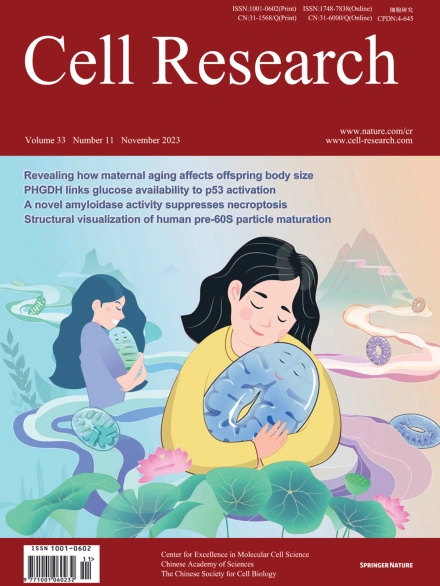
Low glucose metabolite 3-phosphoglycerate switches PHGDH from serine synthesis to p53 activation to control cell fate
Yu-Qing Wu1,† , Chen-Song Zhang1,† , Jinye Xiong1 , Dong-Qi Cai1 , Chen-Zhe Wang1 , Yu Wang1,2 , Yan-Hui Liu1 , Yiming Li2 , Jian Wu2 , Jianfeng Wu3 , Bin Lan4 , Xuefeng Wang4 , Siwei Chen1 , Xianglei Cao1 , Xiaoyan Wei1 , Hui-Hui Hu1 , Huiling Guo1 , Yaxin Yu1 , Shu-Yong Lin1 , Hai-Long Piao5 , Jianyin Zhou2 , Sheng-Cai Lin1,*
1State Key Laboratory of Cellular Stress Biology, Innovation Center for Cell Signaling Network, School of Life Sciences, Xiamen University, Xiamen, Fujian, ChinaGlycolytic intermediary metabolites such as fructose-1,6-bisphosphate can serve as signals, controlling metabolic states beyond energy metabolism. However, whether glycolytic metabolites also play a role in controlling cell fate remains unexplored. Here, we find that low levels of glycolytic metabolite 3-phosphoglycerate (3-PGA) can switch phosphoglycerate dehydrogenase (PHGDH) from cataplerosis serine synthesis to pro-apoptotic activation of p53. PHGDH is a p53-binding protein, and when unoccupied by 3-PGA interacts with the scaffold protein AXIN in complex with the kinase HIPK2, both of which are also p53-binding proteins. This leads to the formation of a multivalent p53-binding complex that allows HIPK2 to specifically phosphorylate p53-Ser46 and thereby promote apoptosis. Furthermore, we show that PHGDH mutants (R135W and V261M) that are constitutively bound to 3-PGA abolish p53 activation even under low glucose conditions, while the mutants (T57A and T78A) unable to bind 3-PGA cause constitutive p53 activation and apoptosis in hepatocellular carcinoma (HCC) cells, even in the presence of high glucose. In vivo, PHGDH-T57A induces apoptosis and inhibits the growth of diethylnitrosamine-induced mouse HCC, whereas PHGDH-R135W prevents apoptosis and promotes HCC growth, and knockout of Trp53 abolishes these effects above. Importantly, caloric restriction that lowers whole-body glucose levels can impede HCC growth dependent on PHGDH. Together, these results unveil a mechanism by which glucose availability autonomously controls p53 activity, providing a new paradigm of cell fate control by metabolic substrate availability.
https://doi.org/10.1038/s41422-023-00874-4